Week 6: Jada Okaikoi
Jada Okaikoi | Clinician Mentor: Dr. Jiwon Kim
This week, I continued working on my research project by segmenting the right ventricular wall from cardiac CT scans taken before and after tricuspid regurgitation interventions. I used 3D Slicer, a medical imaging software that was new to me, to import the scans and create the segmentations (Fig 1.). This involved identifying the boundaries of the right ventricular wall across multiple image slices and manually correcting areas where the wall was difficult to distinguish from surrounding tissue. After completing the segmentations, I performed post-processing steps on the 3D reconstructions, including smoothing the surfaces and checking for areas that required additional correction.
I also began learning the fundamentals of finite element analysis through an introductory tutorial (https://youtu.be/GHjopp47vvQ?si=ADEVmv_QD2cZL2io). I focused on how a 3D model is divided into smaller elements to create a mesh and how material properties, forces, pressures, and boundary conditions are applied. These inputs can then be used to calculate deformation, stress, and strain throughout the model. Understanding these steps will help prepare me to use the reconstructed right ventricular models to compare ventricular mechanics before and after tricuspid regurgitation interventions.
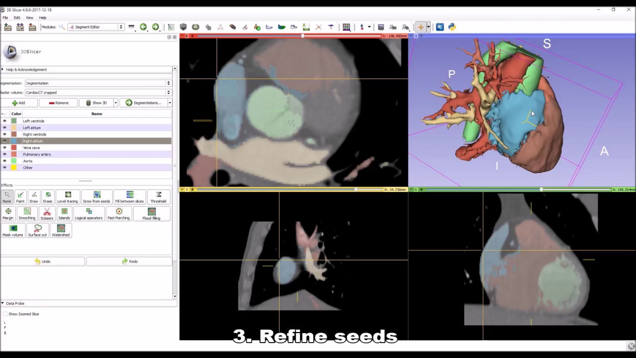
Fig 1. Example of 3D segmentation of the heart in 3D Slicer (https://i.ytimg.com/vi/BJoIexIvtGo/maxresdefault.jpg)


Comments
Post a Comment